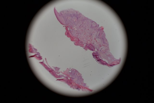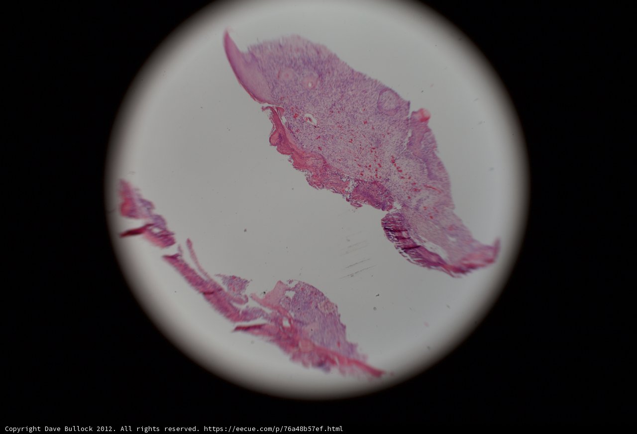
Stained Tissue Micrograph
Monday, July 23rd, 2012 at 1:00:40 PM · Photo Archive/2012/photo_micrographs
A close up of a pink and white tissue, stained and captured through a microscope in 2012.
Chronologically Adjacent
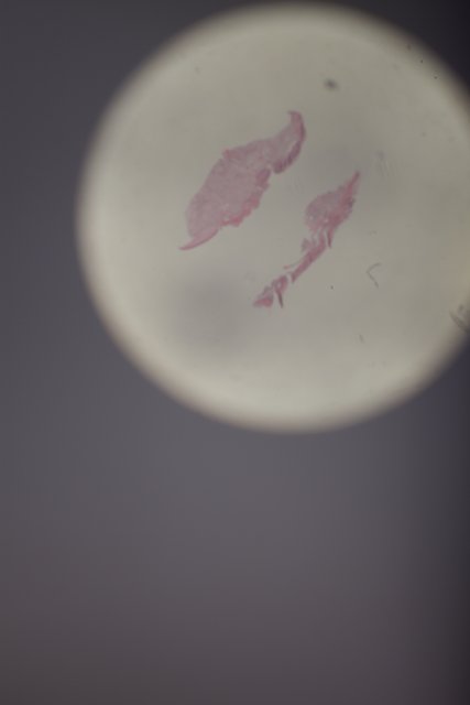
Monday, July 23rd, 2012 at 1:00:40 PM · Photo Archive/2012/photo_micrographs
A close up of a pink and white tissue, stained and captured through a microscope in 2012.
Chronologically Adjacent